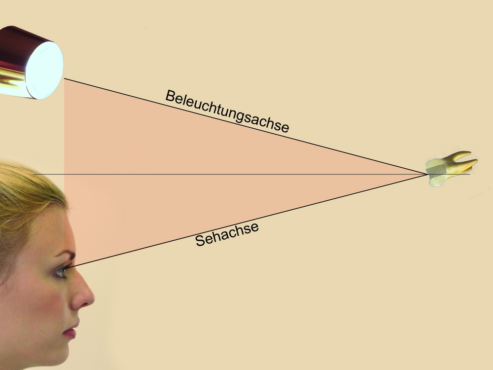
The problem of illuminationIn conventional surgical luminaires, the illumination axis is at an angle to the visual axis. Constant care must be taken to ensure that the light reflected from the object being viewed falls in the direction of the visual axis into the eye of the observer. This can be done either by correcting the position of the operating light or that of the head. In addition, the illumination axis must be free of obstructions so that no shadows are created. In the case of convex formations, a reasonably shadow-free image is possible with a lot of effort (first image). With concave examination areas, e.g. a trephination opening of a root canal treatment, this is almost impossible (second image). |
|

|
|
The solution: coaxial lightingWith coaxial lighting, the visual axis and the illumination axis ideally coincide. The result is a shadow-free, bright image. The light source is located between the examiner's eyes. What the examiner sees is therefore automatically bright; it is not necessary to adjust the light source. |
|